Research Article - Archives of Clinical and Experimental Surgery (2023)
Management of Choledochal Cyst at Tertiary Centre: A Review of Literature and Personal Experience
Rakesh R1*, Rambhupal Choudary2, Prashant Kanni Y3, Praveen Mathew3, Sidhartha Naidu B3 and Reshma Rajeev32Department of General Surgery, Vydehi Institute of Medical Sciences and Research Centre, Bangalore, India
3Department of Medical Gastroenterology, Vydehi Institute of Medical Sciences and Research Centre, Bangalore, India
Rakesh R, Department of Surgical Gastroenterology, Vydehi Institute of Medical Sciences and Research Centre, Bangalore, India, Tel: +919535083450, Email: rakeshreddy.r29@gmail.com
Received: 29-Nov-2023, Manuscript No. EJMACES-23-118439; Editor assigned: 30-Nov-2023, Pre QC No. EJMACES-23-118439 (PQ); Reviewed: 15-Dec-2023, QC No. EJMACES-23-118439; Revised: 22-Dec-2023, Manuscript No. EJMACES-23-118439 (R); Published: 29-Dec-2023
Abstract
Background: Choledochal cysts are dilations of the bile ducts, occurring in both children and adults. In adults, the clinical course differs due to associated hepatobiliary disease. Cysto-enterostomy was the primary treatment but had limitations, so complete cyst excision is preferred when possible to avoid recurrence and malignant change.
Method: A 5-year retrospective analysis was conducted on 33 patients diagnosed with choledochal cysts and treated at the Department of General Surgery and Surgical Gastroenterology between January 2015 and February 2020.
Results: The study involved 33 patients with choledochal cysts, predominantly females (63%), with an average age of 28 years. Abdominal pain and vomiting were the most common symptoms. The Todani classification system was used to categorize the cysts, with the majority being type I (51.5%) and IV A (36.3%). 18% of cases had Abnormal Pancreatico Biliary Junction (APBJ) abnormality and only one patient had raised pre-operative CA-19.9 levels (Cyst Excision). Majority of patients (31) underwent complete cyst excision and HJ (Hepatico Jejunostomy), with only one anastomotic leak and one patient developing pleural effusion and pulmonary embolism. All specimens were suggestive of choledochal cysts with no malignant features. One patient developed anastomotic stricture with hepatolithiasis on follow-up and underwent re-do HJ.
Conclusion: Management of choledochal cyst involves detailed evaluation preoperatively with Magnetic Resonance Cholangio Pancreatography (MRCP) to categorise the type of cyst and to plan the surgery which involves complete excision of cyst with reconstruction by hepatico-jejunostomy. Complete excision of cyst gives a good outcome with minimal morbidity. Long term follow up is essential to detect and manage complications and malignant change.
Keywords
Choledochal cyst; Hepaticojejunostomy; Bile duct; Biliary tree
Introduction
Choledochal cysts, though infrequent, pose a distinctive challenge across pediatric and adult populations. Their precise etiology remains elusive, with a prevailing theory suggesting an abnormal union between pancreatic and biliary ducts, particularly a common channel near the ampullary sphincter—a phenomenon termed Anomalous Pancreatico Biliary Ductal Union (APBDU). Experimental evidence linking APBDU to choledochal cysts supports this theory, though its inconsistent occurrence in affected individuals necessitates further exploration [1,2].
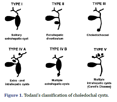
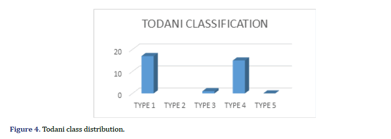
In clinical practice, the Todani classification system, founded on anatomical characteristics and biliary involvement, is widely embraced. Choledochal cyst classified into 5 types, Type I choledochal cysts are subclassified into three types, Type-IA cystic dilation of the extrahepatic duct. Type-IB focal segmental dilation of the extrahepatic duct. Type-IC fusiform dilation of the entire extrahepatic bile duct. Type-II simple diverticula of the common bile duct. Type-III cyst/choledochocele distal intramural dilation of the common bile duct within the duodenal wall. Type-IVA combined intrahepatic and extrahepatic duct dilation. Type-IVB multiple extrahepatic bile duct dilations. Type-V/Caroli disease multiple intrahepatic bile duct dilation. Noteworthy is the prevalence of type I and type IV cysts, influencing study findings significantly [3-6].
The presentation of choledochal cysts manifests variably, with symptoms including abdominal pain, jaundice, and complications such as infection and pancreatitis. The age at presentation correlates with cyst type and severity, with most cases diagnosed in infancy or childhood. Timely diagnosis, regular follow-up, and surgical intervention are paramount to prevent complications. The primary treatment goal is complete cyst removal and biliary reconstruction, with the specific surgical approach dictated by cyst location and extent. Common procedures encompass cystectomy and partial resection with biliary reconstruction when complete removal is challenging [7- 10].
This paper provides a comprehensive review of the current understanding of choledochal cysts in adults, accentuating diagnosis, classification, and tailored management strategies. The rarity of the condition underscores the importance of continued research to enhance our understanding of underlying mechanisms and improve diagnostic and therapeutic approaches. By exploring the intricacies of choledochal cysts, this review contributes to the broader medical knowledge base, guiding clinicians in providing optimal care for affected individuals. Through ongoing research, we aim to refine our comprehension of this enigmatic condition and advance towards more effective clinical management strategies (Figure 1).
Materials and Methods
In this retrospective study, a comprehensive analysis was conducted on 33 patients diagnosed with choledochal cysts who underwent treatment at the Department of General Surgery and Surgical Gastroenterology of Vydehi Institute of Medical Sciences and Research Centre. The study spanned a five-year period, from January 2017 to February 2022.
Patient records were systematically reviewed, encompassing diagnostic information, imaging results, surgical procedures, and postoperative outcomes. This thorough examination aimed to provide detailed insights into the clinical history and management of choledochal cyst cases within the institution. The Todani classification system was utilized to categorize choledochal cysts based on anatomical characteristics and the degree of biliary involvement, facilitating a nuanced understanding of the diverse spectrum of cyst types encountered in the patient cohort.
The clinical presentation of choledochal cysts was investigated, with emphasis placed on various symptoms and complications observed during the study period. This exploration aimed to enhance understanding of how choledochal cysts manifest in patients.
Detailed insights into surgical interventions performed on the patients were documented. This involved a meticulous examination of procedures such as cystectomy and biliary reconstruction, shedding light on the specific approaches employed in the management of choledochal cysts. Postoperative outcomes, including recovery and any complications, were carefully documented to assess the efficacy of the treatment strategies employed. This evaluation contributes valuable information to our understanding of the overall success and challenges associated with managing choledochal cysts.
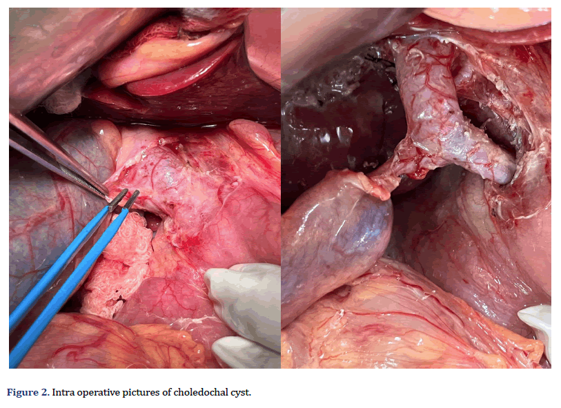
By focusing on a specific patient cohort within the Vydehi Institute of Medical Sciences and Research Centre, this study adds a localized perspective to the existing body of knowledge on choledochal cysts. The findings not only contribute to the institution’s understanding of the condition but also provide insights that may inform broader practices in the diagnosis and management of choledochal cysts. This retrospective analysis serves as a valuable resource for clinicians, researchers, and medical professionals involved in the care of patients with choledochal cysts (Figure 2).
Results and Discussion
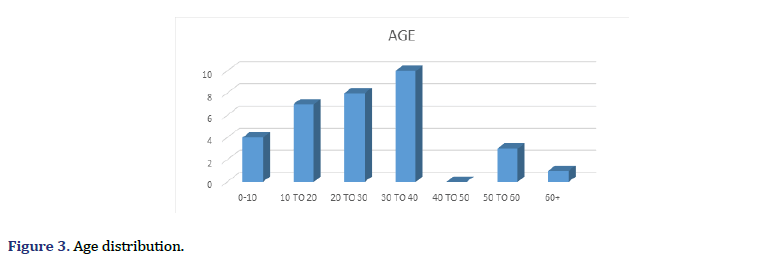
In this investigation, 33 patients diagnosed with choledochal cysts were scrutinized, with a predominant representation of females (63%) and an average age of 28 years. The primary symptoms reported included abdominal pain and vomiting. Employing the Todani classification system, the researchers categorized the cysts, revealing that the majority were type I (51.5%) and IV A (36.3%). An intriguing finding was the identification of an abnormality known as APBJ (anomalous pancreatico-biliary junction) in 18% of cases, while only one patient exhibited elevated pre-operative CA-19.9 levels, a tumor marker.
The predominant surgical approach involved complete cyst excision and hepaticojejunostomy, performed in the majority of patients (31 out of 33). Complications were relatively low, with only one reported case of anastomotic leak and another patient experiencing pleural effusion and pulmonary embolism. Importantly, all excised specimens exhibited characteristics consistent with choledochal cysts, and none displayed malignant features.
During follow-up, one patient developed anastomotic stricture coupled with hepatolithiasis, necessitating a re-do hepaticojejunostomy. This underscores the critical importance of long-term monitoring and management in choledochal cyst cases.
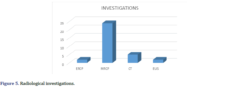
In summary, the study provides valuable insights into the clinical presentation, surgical management, and outcomes of patients with choledochal cysts. The low incidence of malignant features in excised specimens and the successful application of hepaticojejunostomy as a treatment modality highlight the effectiveness of the chosen surgical approach in this cohort. However, the occurrence of complications and the need for a re-do procedure emphasize the ongoing challenges and complexities associated with the management of choledochal cysts. These findings contribute to our understanding of optimal treatment strategies and underscore the importance of vigilant postoperative care in improving overall patient outcomes (Figures 3-6).
Conclusion
In conclusion, the proficient management of choledochal cysts necessitates a meticulous preoperative evaluation, wherein Magnetic Resonance Cholangio Pancreatography (MRCP) assumes a pivotal role in delineating the cyst’s characteristics and guiding the formulation of a tailored surgical strategy. MRCP stands out as an indispensable imaging modality, precisely identifying the choledochal cyst type and furnishing crucial insights for nuanced surgical interventions.
The established protocol for choledochal cyst management encompasses the thorough excision of the cyst, complemented by hepaticojejunostomy reconstruction. This methodical approach not only yields favorable immediate outcomes but also mitigates adverse health repercussions. The paramount importance of a comprehensive preoperative assessment, especially through MRCP, cannot be overstressed, as it serves as a linchpin in tailoring surgical interventions to the specific choledochal cyst subtype.
The chosen procedure, entailing complete cyst removal and hepaticojejunostomy, aligns seamlessly with the observed positive outcomes in the study, thereby contributing to the overall success of the management approach. Furthermore, the emphasis on longterm monitoring emerges as a critical component in the holistic care of individuals with choledochal cysts. The proactive identification and timely resolution of complications, exemplified by reported cases of anastomotic stricture and hepatolithiasis, underscore the importance of continuous surveillance to ensure sustained patient well-being.
In light of these compelling findings, it is incontrovertible that the amalgamation of a thorough preoperative assessment, precise surgical intervention, and vigilant postoperative monitoring constitutes a comprehensive and efficacious strategy for choledochal cyst management. The spotlight on complete cyst removal not only guarantees positive immediate outcomes but also diminishes the risk of enduring health complications, including malignancy development.
This integrated strategy holds promise for optimizing patient outcomes, reflecting the evolving landscape of choledochal cyst management.
References
- Soares KC, Arnaoutakis DJ, Kamel I, Rastegar N, Anders R, Maithel S, et al. Choledochal cysts: Presentation, clinical differentiation, and management. J Am Coll Surg 2014; 219(6):1167.
[Crossref] [Google Scholar] [Pubmed]
- Basaranoglu M, C Balci N, Klor HU. Anomalous Pancreatico Biliary Junction (APBJ) with the drainage of the uncinate process into the minor papilla: Demonstration by MRI. Br J Radiol 2005 ;78(931):655-658.
[Crossref] [Google Scholar] [Pubmed]
- Park DH, Kim MH, Lee SK, Lee SS, Choi JS, Lee YS, et al. Can MRCP replace the diagnostic role of ERCP for patients with choledochal cysts?. Gastrointest Endosc 2005;62(3):360-366.
[Crossref] [Google Scholar] [Pubmed]
- Edil BH, Cameron JL, Reddy S, Lum Y, Lipsett PA, Nathan H, et al. Choledochal cyst disease in children and adults: A 30-year single-institution experience. J Am Coll Surg 2008;206(5):1000-1005.
[Crossref] [Google Scholar] [Pubmed]
- Sacher VY, Davis JS, Sleeman D, Casillas J. Role of magnetic resonance cholangiopancreatography in diagnosing choledochal cysts: Case series and review. World J Radiol 2013;5(8):304.
[Crossref] [Google Scholar] [Pubmed]
- Todani T, Watanabe Y, Narusue M, Tabuchi K, Okajima K. Congenital bile duct cysts: Classification, operative procedures, and review of thirty-seven cases including cancer arising from choledochal cyst. Am J Surg 1977;134(2):263-269.
[Crossref] [Google Scholar] [Pubmed]
- Brown ZJ, Baghdadi A, Kamel I, Labiner HE, Hewitt DB, Pawlik TM, et al. Diagnosis and management of choledochal cysts. HPB 2023;25(1):14-25.
[Crossref] [Google Scholar] [Pubmed]
- Jesudason SB, Jesudason MR, Mukha RP, Vyas FL, Govil S, Muthusami JC, et al. Management of adult choledochal cysts–a 15-year experience. HPB 2006;8(4):299-305.
[Crossref] [Google Scholar] [Pubmed]
- Ando H, Kaneko K, Ito T, Watanabe Y, Seo T, Harada T, et al. Complete excision of the intrapancreatic portion of choledochal cysts. J Am Coll Surg 1996;183(4):317-321.
[Google Scholar] [Pubmed]
- Serin KR, Ercan LD, Lbis C, Ozden I, Tekant Y. Choledochal cysts: Management and long-term follow-up. Surgeon 2021;19(4):200-206.
[Crossref] [Google Scholar] [Pubmed]
Copyright: © 2023 The Authors. This is an open access article under the terms of the Creative Commons Attribution NonCommercial ShareAlike 4.0 (https://creativecommons.org/licenses/by-nc-sa/4.0/). This is an open access article distributed under the terms of the Creative Commons Attribution License, which permits unrestricted use, distribution, and reproduction in any medium, provided the original work is properly cited.
