Case Report - Archives of Clinical and Experimental Surgery (2023)
Successful Laparoscopic Management of Pericaecal Hernia Causing Small Bowel Obstruction
Hans Mare* and William Edward TjhinHans Mare, Department of General Surgery, Rockingham General Hospital, Western Australia, Australia, Tel: +610420780546, Email: hansmare01@gmail.com
Received: 26-Dec-2022, Manuscript No. EJMACES-23-84651; Editor assigned: 28-Dec-2022, Pre QC No. EJMACES-23-84651 (PQ); Reviewed: 13-Jan-2023, QC No. EJMACES-23-84651; Revised: 20-Jan-2023, Manuscript No. EJMACES-23-84651 (R); Published: 30-Jan-2023
Abstract
Background: Small bowel obstruction as a surgical condition can vary greatly in its management. While most cases can be successfully treated conservatively, a not- insignificant number of patients require surgical intervention, particularly those with internal hernias as the cause of their obstruction. Due to the nature of small bowel obstructions often leading to significant abdominal distension, laparoscopic intervention has been avoided to reduce the risk of perforating loops of bowel on entry, with laparotomy being the favoured procedure as a result.
Case presentation: A 54-year-old woman presented with symptoms and signs suggestive of a small bowel obstruction. She had a computed tomography scan that effectively confirmed a closed loop small bowel obstruction in her right lower quadrant. Her abdominal distension improved following nasogastric decompression and was therefore deemed suitable for a diagnostic laparoscopy rather than laparotomy. On entry a pericaecal hernia was noted and extensive adhesiolysis performed to successfully reduce the small bowel contained within. No bowel resection was performed, and the patient was discharged home on day 3 following her procedure.
Conclusion: Laparoscopic surgery is a viable alternative to laparotomy in patients who require operative intervention for acute small bowel obstruction secondary to internal hernia without significant abdominal distension.
Keywords
Small bowel obstruction; Pericaecal hernia; Internal hernia; Laparoscopic surgery
Introduction
Small bowel obstruction is the most common manifestation of an internal hernia in adults. Of the different types of internal hernias, pericaecal are exceedingly rare and usually managed through an emergency laparotomy. We present the case of a 54 year old female who presented with small bowel obstruction secondary to a pericaecal hernia that was successfully treated through laparoscopy.
Case Presentation
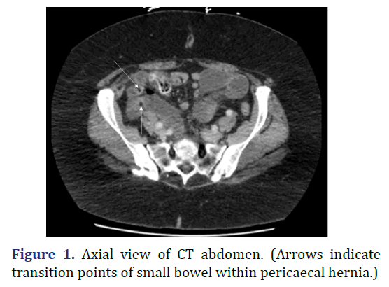
A 54-year-old woman presented to the emergency department with a one-day history of colicky right iliac fossa pain with migration to her epigastrium. Pain was associated with vomiting and exacerbated with oral intake. The patient had previously undergone an appendicectomy, cholecystectomy, gastric band insertion and removal as well as a sleeve gastrectomy, all laparoscopic procedures. On examination in the emergency department the patient’s observations were within normal limits. A soft, distended abdomen was noted with tenderness in the right iliac fossa. A Computed Tomography (CT) scan demonstrated a closed loop small-bowel obstruction with multiple transition points around a single loop in the right lower quadrant, lateral to the caecum (Figure 1). A nasogastric tube was inserted as soon as the diagnosis was made in order to decompress the abdomen, increase intraperitoneal space and reduce the risk of bowel injury intraoperatively. The patient then proceeded to diagnostic laparoscopy.
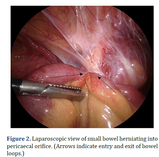
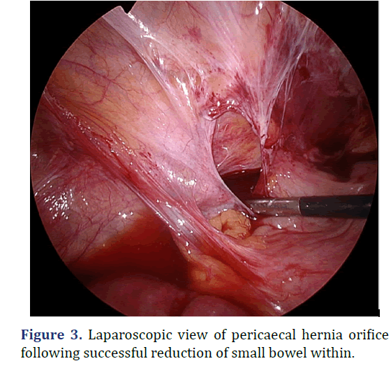
Laparoscopic entry was achieved through the infraumbilical virgin part of the abdomen. A right colon adhesion plate to the right lateral abdominal wall with a small, tight opening around the caecum was found to have created a cocoon through which a 20 cm length of mid small bowel herniated (Figure 2). This opening was widened and extensive adhesiolysis performed without the use of an energy device (Figure 3). An a-trac laparoscopic grasper was used on the mesentery instead of the bowel during reduction. The bowel contained within was initially dusky, however a return of colour and vigour was observed after pause. A small bowel resection was not performed. The patient had an uncomplicated recovery and was discharged home day 3 post-operatively following gradual upgrade of oral dietary intake.
This patient’s history and examination were suggestive of a small bowel obstruction. While her CT scan was able to confirm the diagnosis of a closed loop obstruction, it did not detect the presence of a pericaecal internal hernia. Internal hernias are defined as a protrusion of a viscus, usually small bowel, through a peritoneal or mesenteric aperture within the abdominal cavity. Internal hernias are uncommon and typically manifest with a small bowel obstruction. Approximately 0.6%-5.8% of small bowel obstructions in adults can be attributed to internal hernias [1], and pericaecal hernias are rarer still, constituting up to only 6%-13% of all internal hernias [2]. Closed loop small bowel obstructions that occur from internal hernias can rapidly progress from incarceration to strangulation and full thickness bowel necrosis [3].
Results and Discussion
Pericaecal hernias emerge through an aperture that develops within the peritoneal recess formed by local folds of the peritoneum [4]. Four subtypes of pericaecal hernias have been described based on the anatomic location of the recess including ileocolic, retrocolic, ileocaecal and paracaecal [5]. Most commonly the loop of herniated bowel consists of an ileal segment protruding through a defect in the caecal mesentery and extending into the right paracolic gutter [2]. Patients with pericaecal hernias present in a manner similar to those with other types of internal hernias, except for the location of symptoms which is typically in the right lower quadrant, therefore pericaecal hernias are occasionally mistaken for appendiceal abnormalities [5].
Laparotomy has long been the standard operative approach to managing acute small bowel obstructions, particularly those due to internal hernias. Due to the difficulty in establishing a working space, visualising the site of obstruction and risk of injury to the distended bowel, laparoscopy for small bowel obstructions was previously deemed inappropriate [6]. There are however increasing numbers of case reports and studies demonstrating success in managing small bowel obstructions secondary to internal hernias through laparoscopy [7]. To our best knowledge this is one of only a few reported cases of a pericaecal internal hernia with small bowel obstruction being managed successfully through laparoscopy. Less than 35 cases of pericaecal hernias have been reported in the literature, with the vast majority being managed through laparotomy [7,8].
Conclusion
While rare, the consideration of a pericaecal hernia as a diagnosis is important in the patient presenting with right lower quadrant pain, particularly when signs and symptoms of small bowel obstruction are present. We believe this case successfully demonstrates the utility of CT as a diagnostic tool and the practicality of laparoscopic surgery in the management of pericaecal hernias causing small bowel obstruction.
Declarations
Conflicts of interest
Authors have no conflicts of interest to declare.
Ethics statements
This case report briefly reviews the relevant literature surrounding the successful use of a common surgical technique in treatment of small bowel obstruction with an internal hernia. All details that might disclose the identity of the subject under study for this case report has been omitted.
The patient who’s case is being presented here gave their consent to have their case written up and published in a relevant journal.
Consent for publication
The work presented here has not been published before and is not under consideration for publication elsewhere. The publication has been approved by all co-authors. No experimentation was performed on human subjects.
References
- Monica ML, Antonella M, Gloria A, Diletta C, Nicola M, Ginevra D, et al. Internal hernias: A difficult diagnostic challenge. Review of CT signs and clinical findings. Acta Bio Med 2019;90(Suppl 5):20.
[Crossref] [Google Scholar] [Pubmed]
- Martin LC, Merkle EM, Thompson WM. Review of internal hernias: Radiographic and clinical findings. AJR Am J Roentgenol 2006;186(3):703.
[Crossref] [Google Scholar] [Pubmed]
- Paulson EK, Thompson WM. Review of small-bowel obstruction: The diagnosis and when to worry. Radiology 2015;275(2):332-42.
[Crossref] [Google Scholar] [Pubmed]
- Rivkind AI, Shiloni E, Muggia-Sullam M, Weiss Y, Lax E, Freund HR, et al. Paracecal hernia: A cause of intestinal obstruction. Dis Colon Rectum 1986;29(11):752-4.
[Crossref] [Google Scholar] [Pubmed]
- Mathieu D, Luciani A. Internal abdominal herniations. AJR Am J Roentgenol 2004;183(2):397-404.
[Crossref] [Google Scholar] [Pubmed]
- O’Connor DB, Winter DC. The role of laparoscopy in the management of acute small-bowel obstruction: A review of over 2,000 cases. Surg Endosc 2012;26(1):12-7.
[Crossref] [Google Scholar] [Pubmed]
- Ogami T, Honjo H, Kusanagi H. Pericecal hernia manifesting as a small bowel obstruction successfully treated with laparoscopic surgery. J Surg Case Rep 2016;2016(3).
[Crossref] [Google Scholar] [Pubmed]
- Chia DKA, Tay KV, Kow A, So J, Shabbir A, Kim G, et al. Paracaecal hernia: Uncommon but important cause of small bowel obstruction successfully managed with laparoscopic surgery. ANZ J Surg 2019;89(6):769-70.
[Crossref] [Google Scholar] [Pubmed]
Copyright: © 2023 The Authors. This is an open access article under the terms of the Creative Commons Attribution NonCommercial ShareAlike 4.0 (https://creativecommons.org/licenses/by-nc-sa/4.0/). This is an open access article distributed under the terms of the Creative Commons Attribution License, which permits unrestricted use, distribution, and reproduction in any medium, provided the original work is properly cited.


